Assessment of Myocardial Viability Using Nuclear Medicine Imaging in Dextrocardia
Por um escritor misterioso
Last updated 30 maio 2024
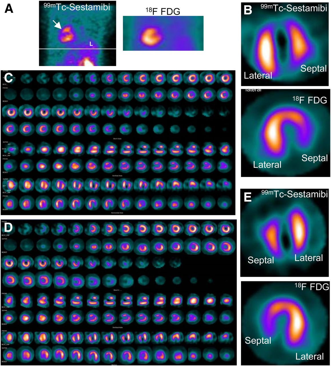
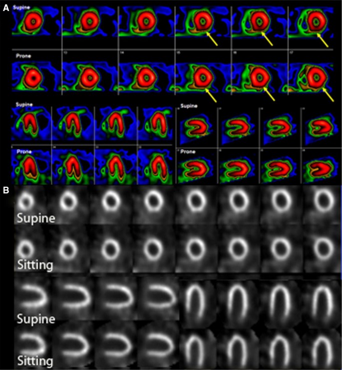
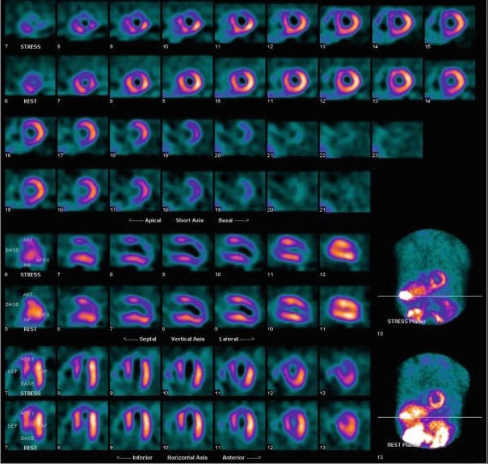
Imaging of dextrocardia in humans requires an understanding of the orientation of the heart chambers and walls. There are many types of cardiac malpositioning, such as dextrocardia (with or without situs inversus), mesocardia, and levocardia. Myocardial perfusion scintigraphy of dextrocardia has been explained in case reports and imaging atlases; however, myocardial viability assessment using nuclear medicine imaging techniques is less documented in the literature. Methods: In 2 cases of dextrocardia with situs inversus and 1 case of mesocardia, myocardial viability was assessed using 99mTc-sestamibi rest perfusion scintigraphy and 18F-FDG PET. Cardiac SPECT images of dextrocardia with situs inversus were acquired using the feet-first supine position with a 180° arc from left anterior oblique to right posterior oblique, whereas a right-lateral–to–left-lateral arc was used for mesocardia. The processing and reconstruction were done by entering the dataset for the feet-first supine position and repeating after entering the dataset for the feet-first prone position. The 2 sets of reconstructed images were compared for orientation of walls and cardiac chambers. Results: The first processing, using the feet-first supine position, revealed an interchanged septum and lateral wall in reconstructed images of dextrocardia with situs inversus. This interchange was corrected by changing the position to prone during processing of the rest perfusion and PET raw data. The display of cardiac slices in various axes matched the conventional nomenclature for the septum and lateral wall, leading to easy interpretation. However, this change was not required in the mesocardia, for which the location of the heart chambers was not interchanged. Conclusion: Because the acquisition protocol for SPECT is a semicircular orbit, the various types of dextrocardia require careful selection of the arc, with the patient positioning kept feet-first supine. Processing and reconstruction of data by changing the patient position to prone was found to be most useful method of matching the septum and lateral wall orientation for interpretation of images.
Incidental Findings on Myocardial Perfusion SPECT Images
Society for Cardiovascular Magnetic Resonance/European Society of

SPECT myocardial perfusion imaging in patients with Dextrocardia
Assessing Myocardial Viability in Clinical Practice - ABC Imaging
Single Photon Emission Computed Tomography (SPECT) Myocardial
Incidental Findings on Myocardial Perfusion SPECT Images
Nuclear Imaging in Stable Ischemic Coronary Disease

Society for Cardiovascular Magnetic Resonance/European Society of
Assessment of Myocardial Viability in Patients with Heart Failure
Recomendado para você
-
 Brain Test Level 372 He wants big muscles Walkthrough30 maio 2024
 -
 how to solve brain test 372|TikTok Search30 maio 2024
 -
 Easy Game Brain Test Level 372 Finish shopping.30 maio 2024
 -
 Cube Meet APK for Android Download30 maio 2024
 -
 A sense of self30 maio 2024
 -
 Brain Test Level 372 solução dos jogos #braintest #respostas #shorts #game #jogos30 maio 2024
 -
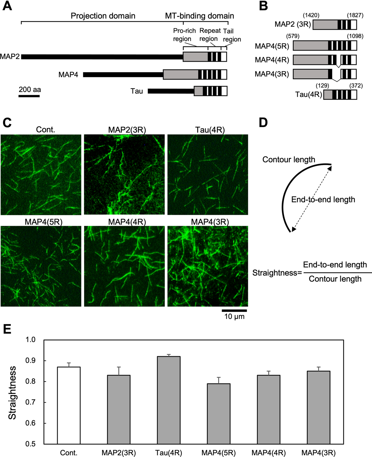 Effects of three microtubule-associated proteins (MAP2, MAP4, and Tau) on microtubules' physical properties and neurite morphology30 maio 2024
 -
 Selamatkan kapal Titanic! (Brain Test Level 372) - CadeMedia30 maio 2024
 -
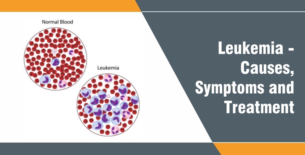 Blood Cancer - Causes, Symptoms and Treatment - Rela Hospital30 maio 2024
 -
 How to Block Spam Emails30 maio 2024
você pode gostar
-
 Roblox Grand Piece Online: Magu Magu no mi(Magma Magma Devil Fruit30 maio 2024
 -
 Fnaf 3 Animatronic Minigames 2 by Spring-o-bonnie on DeviantArt30 maio 2024
 -
 Razones para rechazar el proyecto del casino online en Córdoba - ENREDACCIÓN - Córdoba - Argentina30 maio 2024
 -
Pelé” foi adicionado ao dicionário de português como um adjetivo. O n30 maio 2024
-
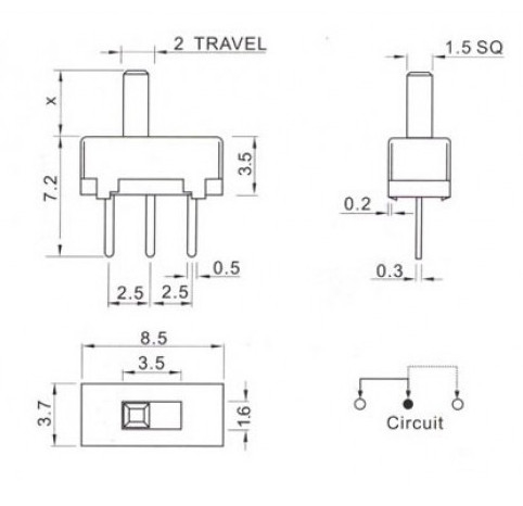 Chave liga-desliga reversível para protoboard30 maio 2024
 -
 o que ha em jupiter brain test30 maio 2024
 -
:max_bytes(150000):strip_icc()/Stranger-Things-recap_004-3dc82704580e4734a727fd0afdccabec.jpg) Stranger Things season 4, Vol. 1 episodes 1-7 recap30 maio 2024
Stranger Things season 4, Vol. 1 episodes 1-7 recap30 maio 2024 -
 Putin meets Russia's national chess team before 43rd Chess Olympiad in Georgia30 maio 2024
 -
 Quebra cabeça importado Grafika - Trente Glorieuses - 3000 peças30 maio 2024
 -
 Nintendo Switch ROMs: Guide for Download and Play30 maio 2024
